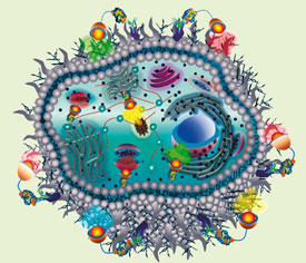
Vasudevanpillai Biju and colleagues at National Institute of Advanced Industrial Science and Technology (AIST) in Japan look at how nanoparticles might light up cells to unravel concealed subcellular structures and biomolecular functions.
The brilliant colours of nanoparticles have attracted biologists and biomedical researchers to unconventional bioimaging since the late 1990s. Nanoparticles have the power to literally light up concealed structures and vital functions in cells, and have replaced organic dyes used for this purpose. In particular, size-dependent tunable photoluminescence colour and exceptional photostability of semiconductor quantum dots (QDs) has brought radical changes to modern biomolecular and cell imaging. As such, QDs are the smartest pigments in the colour box of modern biologists and biomedical researchers.
Material chemists have surprised biologists with their ability to customise organometallic reactions for the nucleation and growth of assorted multicolour to size-selected monochromatic QDs from various combinations among transition metals and chalcogens. The most attractive QDs are hydrophobic-capped CdSe and CdTe.
High quality QDs are commonly synthesised in the organic phase and have a highly hydrophobic surface, which twisted biologists' arm to challenge random mixing of QDs with cell and tissue samples. But, synthesised QDs are less accessible to biomolecules, more likely to release toxic ions, and susceptible to photobleaching, so their use in bioimaging has been hindered. Now, stable QDs can be prepared routinely using ZnS shells, allowing tethering of hydrophilic thiols, silanes or polymers, and preparation of biocompatible QDs.

Nanoparticles aid biomedical research in a wide variety of applications
|
Conversion of biocompatible QDs into bioconjugates is a prerequisite for labelling cells. The biomolecule added depends on the particular application aimed for, such as extracellular labelling, intracellular delivery, intracellular labelling, or in vivo imaging.
Cells can be labelled using QDs in a nonspecific or targeted manner. Hydrophobic and electrostatic interactions between capping molecules in QDs and biomolecules in cells induce passive nonspecific labelling. Active nonspecific labeling using QDs is done by transiently destabilising cell membranes using techniques such as nanoinjection, electroporation, or osmotic lysis. But, such methods are not promising for labelling large numbers of cells without affecting cell physiology.
Targeted labelling of cells has been extensively investigated to find a bridge between QDs and extracellular proteins or subcellular organelles using a multitude of antibodies, proteins, peptides, aptamers, nucleic acids, small molecules, and liposomes. Reactive moieties such as streptavidin, biotin, primary amine, thiol, maleimide, succinimide, and carboxylic acids are inevitable supplements in such labelling.
Recently, QDs conjugated with common antibodies and ligands have become commercially available, but customisation is often required to untangle novel problems. While nonspecific approaches have coined a strong interface between biology and nanomaterials. Targeted delivery increasingly offers specific applications such as imaging and photodynamic therapy of cancers. Cancer research has benefitted significantly from advances in QDs and other nanomaterials. QDs assembled with other imaging probes such as magnetic nanoparticles and radio nuclei can be contrast agents for multimodal imaging. Recently, multimodal probes conjugated with antibodies, secondary antibodies and anticancer drugs against breast, prostate, pancreatic, liver, and skin carcinomas are under groundbreaking investigation.
Also, multimodal probes conjugated with photosensitisers might challenge magnetically assisted photodynamic therapy. However, the large hydrodynamic size (50-300 nm) of pre-fabricated probes prevents site-specific delivery and uniform biodistribution. Also, prolonged retention in the immune system and toxicity of multimodal probes hinder the translation of QD technology to biomedical applications. These limitations demand in situ remote fabrication of on-demand degradable probes. In situ near infrared photofabrication of multimodal probes such as fluorescent nanomagnets in cells and tissues might facilitate site-specific delivery and uniform biodistribution. Additionally on-demand photodegradation might detach, fragment and eliminate used probes without allowing prolonged retention in the body.
Read more in the review 'Delivering quantum dots to cells: Bioconjugated quantum dots for targeted and nonspecific extracellular and intracellular imaging' in Chemical Society Reviews


